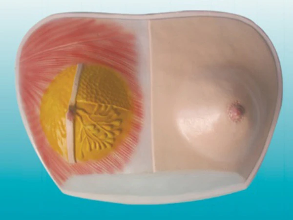
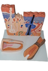
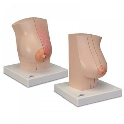
Female chest wall and mammary gland: right mammary gland in lactation with signs of inflammation (mastitis) and left mammary gland out of lactation with signs of various diseases. The model consists of the right female mammary gland in a state of lactation and the surrounding part of the chest wall and the left female mammary gland outside of lactation and the surrounding part of the chest wall.
Both parts of the model are presented in sagittal section. The cut surfaces are represented by the breast tissue as well as the underlying anatomical structures such as muscles, ribs, costal and visceral pleura and lungs.
Inflammation of the mammary gland (mastitis) is represented on the right mammary gland and various other diseases on the left mammary gland. The right mammary gland and chest wall are divided into two halves by a sagittal incision along the median clavicular line and are connected to each other using magnets.
The slice of the right half shows healthy breast tissue, whereas the left half shows changes characteristic...

Tell us what you need and get quotes from verified suppliers