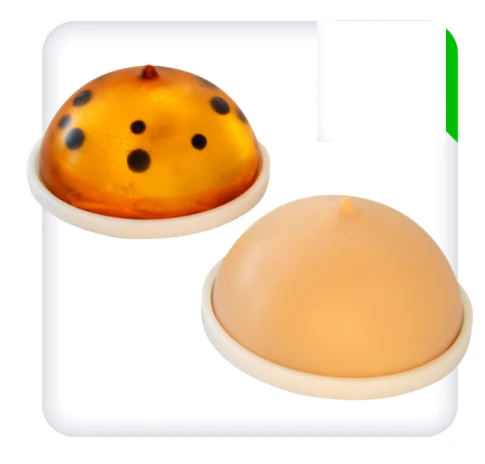
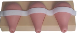
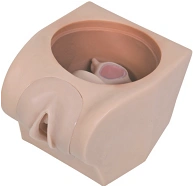
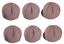
The simulator is designed as a set of mammary gland models placed on the base.
Two types of models are included:
Transparent: initial level, hypoechogenic and hyperechogenic inclusions are stained in different colours. The biopsy is performed under direct visual control.
Opaque: for experienced trainees, covered with imitation skin. The biopsy is performed under the control of an ultrasound machine.
Inside each model there are 12 neoplasms (6 hyperechogenic, 6 hypoechogenic), diameter from 0.6 to 12 mm, located at different levels. The simulator is designed for training in interpretation of information and performing punctures under the control of the ultrasound sensor.
Skills to be practised:
fine-needle aspiration biopsy;
thick-needle biopsy.


Tell us what you need and get quotes from verified suppliers