


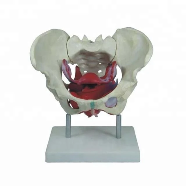
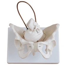
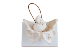
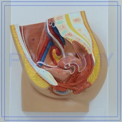
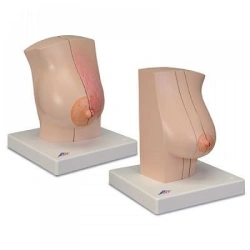
The four-part model of the female pelvis provides detailed information about the topography of the bones, ligaments, pelvic floor muscles and pelvic organs.
The right half of the model shows the pelvic bones with ligaments.
The left half of the pelvic model contains the muscles:
pelvic floor,
including the muscle that lifts the anus,
the sciatic cavernous muscle,
the deep and superficial transverse perineal muscles,
the external sphincter of the anus,
the external sphincter of the urethra.
The vestibular bulb and the large vestibular (Bartholin's) gland can be seen due to the partially detachable bulbous-spongy muscle. The possibility of separation by a median sagittal incision passing through the bladder, vagina, uterus and rectum, the mutual arrangement of the pelvic floor muscles with the openings of the urethra, vagina and rectum can be seen.
Dimensions: 190 x 270 x 190
Weight: 1.75kg.


Tell us what you need and get quotes from verified suppliers