


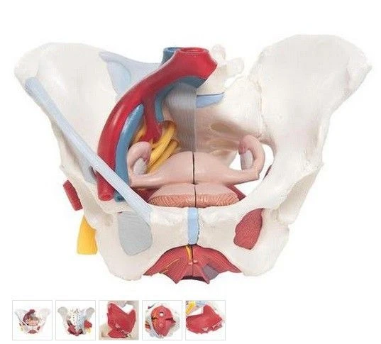
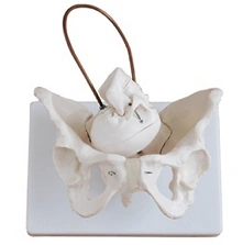
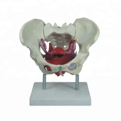
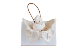
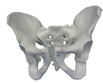
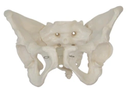
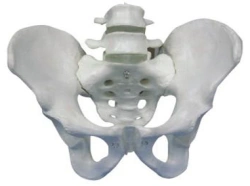
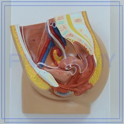
This six-part model of the female pelvis provides detailed information on the topography of the bones, ligaments, pelvic floor muscles and pelvic organs. It represents the entire pelvic floor with the partially detached external sphincter of the anus, the external sphincter of the urethra, the deep and superficial transverse perineal muscles and the bulbous spongiosa muscle cut along the medial sagittal line. The rectum, uterus with fallopian tubes, ovaries and vagina are also separated and can be divided into two parts along the median sagittal incision.
The right half of the pelvic model shows the division and topographic anatomy of the common iliac artery, the external and internal arteries, and the common and external iliac veins. The right sacral plexus, right sciatic nerve, and right genital nerve are also shown.
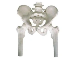
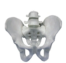
Bones and ligaments represented: two femurs, pubic symphysis, sacrum and coccyx, fifth lumbar vertebra with intervertebral disc. Midline sagittal....

أخبرنا بما تحتاجه واحصل على عروض أسعار من موردين موثوقين